Research Article | | Peer-Reviewed

Effects of Norcantharidin on the Main Organs and Tissues of Mice

Published in Advances (Volume 6, Issue 4)
Received: 26 September 2025     Accepted: 18 October 2025     Published: 24 December 2025
Views:       Downloads:
Abstract

Norcantharidin (NCTD), a demethylated derivative of cantharidin, reportedly exhibits various biological anticancer activities including apoptosis, inhibition of cell proliferation, cell-cycle blockage, induction of cell apoptosis, and anti-angiogenesis. NCTD is currently used as an anticancer drug for hepatoma, breast cancer and colorectal adenocarcinoma. However, the effects of NCTD on the organs and tissues in vivo remain largely unknown. Here, we pathologically examined the effects of NCTD on the organs and tissues including liver, heart, kidney, spleen and medulla in mice. After mice were treated with 60 daily intraperitoneal injections of NCTD (2.5 mg/kg or 5 mg/kg), toxicity on the heart and kidneys were not discovered but on the liver was a significant increase according to the injection dose and the date. The number of leucocytes had a tendency to rise in the peripheral blood test and there weren’t any pathological changes in the spleen and medulla. NCTD has no side effects on spleen and hematopoietic system and applying for a considerable period at a certain dose may not reduce the number of leucocyte but rather shows tendency of increasing which will have great influence on the onco-immune of the host. It is concluded that NCTD is suitable for the application of the anti-cancer treatment with no myelo-suppression.

Published in Advances (Volume 6, Issue 4)
DOI 10.11648/j.advances.20250604.13
Page(s) 123-130
Creative Commons

This is an Open Access article, distributed under the terms of the Creative Commons Attribution 4.0 International License (http://creativecommons.org/licenses/by/4.0/), which permits unrestricted use, distribution and reproduction in any medium or format, provided the original work is properly cited.

Copyright

Copyright © The Author(s), 2025. Published by Science Publishing Group

Keywords

Norcantharidin (NCTD), Toxicity, Anti-tumor Drug

1. Introduction
Cantharidin (CTD) is a naturally occurring compound isolated from the medicinal insect blister beetle (Mylabris phalerata Pallas) . One of the important medicinal uses of CTD is its anti-cancer activities . It can induce p53- dependent apoptosis and double-strand break-age of DNA in cancer cells . CTD treatment causes granulocytosis in vivo but not gran- ulocytopenia due to most chemotherapeutics . Due to these unique biological activities, CTD has a promising compound that can overcome chemical limitations and can be used for cancer treatment. However, the application of CTD has disadvantages that are toxic to the gastrointestinal tract and urinary tract. . From this, the studies to develop a derivative of CTD with less side effects and costs while having its own antitumor activity. Thus, a synthetic of CTD that was developed first is the Norcantharidin (NCTD).
It has been reported that NCTD (NCTD) is a water-soluble synthetic small molecule and a demethylated derivative of cantharidin, which has been shown to exhibit antitumor activity, while being the least toxic to the kidney. Norcantharidin is a potent blocker of protein phosphatase 1 (PP1) and 2A (PP2A), just like CTD, and two phosphatases are known to participate in many different cellular processes, including DNA damage, cell cycle arrest and apoptosis.
NCTD is a heptane-2,3-dicarboxylic anhydride, a demethylated, water-soluble synthetic small molecule of CTD. NCTD has fewer side effects on nephrotoxicity and inflammation than cantharidin . And it is known to have anticancer effects . The cytotoxic and anticancer mechanisms of NCTD are diverse . It can cause cell death, inhibition of angiogenesis, and metastasis in many cell lines and affect several pathways that regulate cell proliferation . Furthermore, NCTD has been shown to inhibit P-glycoprotein (P-gp) and overcome multidrug resistance .
Some researchers have suggested that NCTD is a blocking factor for the activation of Wnt/β-catenin pathways and Wnt target genes such as c-Jun and cyclin D1. The Wnt/β-catenin pathway regulates cell proliferation, migration, apoptosis, differentiation, and stem cell self-renewal .
Chuang et al. noted that NCTD inhibited the proliferation of Jurkat cells by 64% significant β-cateninsignaling in a concentration-dependent manner . In human gallbladder cancer transplant tumors, NCTD-treated group significantly decreased the expression of cyclin-D1 and Bcl-2 and survival proteins/mRNAs . NCTD inhibits the proliferation of gallbladder cancer (GBC-SD) cells, decreasing the expression of proliferation-related genes such as cyclin-D1 or apoptosis-related genes and increasing the rate of cell apoptosis . NCTD also abolishes the cyclin D3, E, A, and B transcripts, thereby arresting cell-cycle progression from G1 to S . Some researchers have suggested that NCTD is an inhibitor of Shh expression in a variety of cell lines and a blocker of Gli-1is nuclear translocation. Hedgehog signaling pathways play an important role as regulators of cell differentiation, tissue polarity, and cell proliferation . Aberrant activation of Hh signaling leads to the formation of CSCs and the development of cancer, and also to cancer angiogenesis and metastatic invasion . Chen et al. demonstrated that NCTD inhibits Shh expression in several cell lines of breast cancer and also blocks the nuclear translocation of Gli-1is .
Besides, the plasma VEGF levels of tumor-bearing mice, migration, and capillary-like tube formation of HUVECs are suppressed by NCTD with potential anti-metastasis and anti-angiogenesis . Also, NCTD inhibits metastasis in CT26 cells by the down- expression of matrix metallo-proteinase-9 (MMP-9) activity .
Recently, we demonstrated that NCTD can be a substrate for P-gp (MDR-1, ABCB1) and can improve multidrug resistance in cancer cells. P-gp acts as a drug efflux pump that pushes a wide range of different chemicals out of MDR cancer cells . It was confirmed that NCTD significantly increased the uptake of lactose-NCTD-conjugated nanoparticles by inhibiting P-gp and multidrug resistance-related protein 2 (MRAP-2) in a heterogeneous human CRC monolayer model . NCTD induces apoptosis of oral cancer cells with resistance to a variety of chemo-anticancer drugs. NCTD has pharmacological potential in the treatment of CSCs .
In summary, the beneficial effects of NCTD may be involved in the regulation of CSC self-renewal pathways, overcoming multidrug resistance, and radiation sensitized effects. Therefore, we injected NCTD into the abdominal cavity of mice and re-examined about the effects on the parenchymal organs and the hematopoietic tissues of mice in order to apply it to the pre-surgical chemotherapy.
2. Materials and Methods
2.1. Materials
Animal model; BALB/c male mice (6–8 weeks old) were obtained from the Laboratory Animal Center in Pyongyang University of Medical Science.
Material; It was diluted with the distilled and sterilized water to use the synthesized and refined Norcantharidin (NCTD) powders in the biochemical laboratory of University of Science.
The purity of the NCTD powders were 99.2% and their analysis results were as follows. {mp: 108~110℃, m/z: 185 [M +H2O-H]-, 1H NMR (100.13MHz, CDCl3, ppm): 3.17 (2H, s, H-c), 5.03 (2H, dd, J=3.0, 3.0Hz, H-b), 1.5~2.2 (4H, m, 2×CH2)}.
The used instruments were High-pressure liquid chromatography (HPLC)-Mass spectrometer (MS) (ACQUITY HPLC SQD-2), Nuclear magnetic resonance (NMR) spectrometer (BRUKER WP 100 SY), Olympus microscope and Digital camera, Micropipettes and so on.
2.2. Methods
2.2.1. Finn Chamber Test-Angiogenesis Test
First by filter paper (nitrocellulose attached on both sides of Polyethylene resin ring model (diameter-10mm, height-2mm) and sterilized. Next the subcultered Croquel-180 Sarcomatous Cells (10day, 5×10⁶/0.1mL) put into its resin ring and closed up. Under the back derma of mice, sterilely made air-pocket (5~7㎖) and incised derma and the resin ring model intered into it. After injecting into abdominal cavity the drugs (NCTD-2.5mg/kg, 5mg/kg; 5-Fu-50mg/kg, 0.9% solution of salt-100㎕) once every day for 3 days or 5 days, next day we observed. We divided the mice (weight: 20-24g) into the experiment groups and control group at 10 per a group without distinction of female and male (Total 60 heads), again the experiment groups into the research-1 group (Inject dose: at NCTD 2.5mg/kg per mouse) and the research-2 group (Inject dose: at NCTD 5mg/kg per mouse), then mice were treated with 60 daily intraperitoneal injections of NCTD. Also, the control group was injected 0.85% salt solution. And, mice were treated at intervals of 30d and compared with the control group.
2.2.2. Body-weight Observation
We divided the mice (weight: 20-24g) into the experiment groups and contrast group at 10 head per a group without distinction of female and male (Total 60 heads), again the experiment groups into the research-1 group (Inject dose: at NCTD 2.5mg/kg per mouse) and the research-2 group (Inject dose: at NCTD 5mg/kg per mouse), then were injected the fixed NCTD into the abdominal cavity of mice once a day for 60d. Also, the contrast group was injected 0.85% salt solution 100μL into the abdominal cavity of mice once a day for 60d. And, the weights of mice were observed at intervals of 30d and compared with the contrast group.
2.2.3. Peripheral Blood Test, Serum Biochemistry Test
As above, while injecting NCTD into the abdominal cavity of mice once a day for 60d, the blood was gathered in the orbital equal pulses of mice, and the number of leukocytes, erythrocytes and thrombocytes were counted by blood testing way at intervals of 30d. Also, the serum transaminase (ALT and AST), the blood urea nitrogen (BUN), the serum creatine (Cre) and the serum colloid reaction test (ZTT) were measured.
2.2.4. Histopathology Test
As above, while injecting NCTD into the abdominal cavity of mice once a day for 60d, the mice were killed by cervical fracture at intervals of 30d and the parenchymal organs and the hematopoietic tissues were extracted and fixed sufficiently in 10% neutral formalin, and went through pathological sample making course, and dyed by H-E stain. Then, we observed about the degeneration and necrosis of the parenchymal cells and appearance of inflammatory cells on an optical microscope of 400 diameters. The 5 regions were confirmed as the most severe changes of the parenchymal cells and were analyzed, and specially were observed the existence of hemorrhage in the kidney. The number of lymph nodules was observed on an optical microscope of 100 diameters in the spleen. Also, the quantity of marrow and the appearance degrees of fatty materials were observed on an optical microscope of 400 diameters in the bone marrow, here this was measured by counting the ratio of area in the cavity and tissues of marrow (Marrow tissues/cavity).
2.3. Ethical Statement
All animal experiments were performed in accordance with the regulations of the Administration of Affairs Concerning Experimental Animals in DPR Korea. The study protocol was approved by the Institutional Animal Care and Use Committee of the Pyongyang University Science and Technology.
3. Result
As the acute toxicity of NCTD (LD50) used in the experiments is 20.75mg/kg, before the aforementioned experiments, we had the experiment for confirming the anti-angiogenesis effects of NCTD below a fourth or a fifth concentration of acute toxicity in chamber-transplant models. In vivo, when NCTD was injected at 2.5mg /kg or 5mg/kg per mouse for 5d, their anti-angiogenesis rates in the fasciae of regions of back were 91.4% or 93.5%, thus they were not significantly different than the anti-angiogenesis rates of 5-Fu (92.1%) of the cases that a dose of 5-fluorouracil (5-Fu) was 10mg/kg (Figure 1). We evaluated on the effects of NCTD affected to the main organs and tissues of mice on the basis of this experimental results.
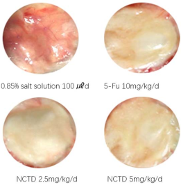
Figure 1. Effect of NCTD on angiogenesis of mice. In figure, in the fasciae of regions of back with the 0.85% salt solution group the blood vessels were abundant and the fasciae of them got so thick too. Also, in the fasciae of regions of back with 5-Fu 10mg/kg/d groups, NCTD 2.5mg/kg/d groups and 5mg/kg/d groups the blood vessels were a little and the fasciae of them don’t get thick. The anti-angiogenesis effect of NCTD was dependent upon a dose of it and expressed from a fixed effective concentration (2.5mg/kg/d) remarkably.
3.1. Effect of NCTD on Liver of Mice
Injecting NCTD at a dose of 2.5, 5mg/kg for 30, 60ds, the value of ALT and AST got increased 0.85% compared to control group and the value of NCTD for 60d were remarkably higher than 30d and injecting at a dose of 5mg/kg showed significant change com- pared to 2.5mg/kg (Figure 2-a, b). Then injecting NCTD at a dose of 2.5 mg/kg for 30, 60d the value of ZTT did not increase compared to control group but at 5mg/kg there was significant increase in the value (Figure 2-c). Also, observing the morphological changes in the liver, after injecting NCTD at a dose of 5mg/kg for 30, 60d there was significant vacuolar degeneration in hepatocytes but at 2.5mg/kg for 30d with less degeneration. The inflammatory cells infiltrated into hepatic tissue as degenerated cells increased in number with more infiltration (Figure 3). Therefore, as the injection dose increases with longer application period the effect of NCTD on the liver would be negative. Also in the case of injecting NCTD at a dose of 2.5mg/kg there weren`t be any difference in the functional and morphological changes in the liver, so if NCTD was injected with low concentration that can show anti-angiogenesis, the effect can be expected without any damages on the liver.
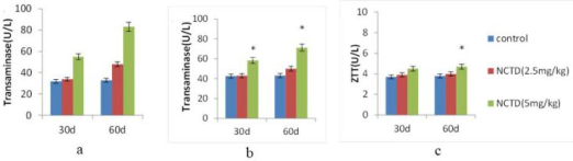
Figure 2. Effect of NCTD on the functional changes of liver of mice. (a, b) The more increased a dose of NCTD and the longer a date of NCTD, the larger the values of transaminase (ALT, AST) were significantly increased. (* p<0.05, versus control) (c) Also, the values of serum colloid reaction test (ZTT) were increased gradually. (* p<0.05, versus control).
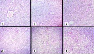
Figure 3. Effect of NCTD on the morphologic changes of liver of mice (H-E stain, 10×40). (a) The normal liver of mice injected at 0.85% salt sodium 100㎕ per mouse. (b) The research liver of mice injected at NCTD 2.5mg/kg once a day for 30d. Here, the hepatocytes had the vacuolar degenerations and the infiltrated inflammatory cells were not found, and this was equal with the control. (c) The research liver of mice injected at NCTD 2.5mg/kg once a day for 60d. The partial hepatocytes in hepatic lobules were become the vacuolar degeneration, and the inflammatory cells were infiltrated as well. (d) The research liver of mice injected at NCTD 5mg/kg once a day for 30d. The partial hepatocytes in hepatic lobules were more severely become the vacuolar degeneration than the former, and the many inflammatory cells were infiltrated too. (e, f) The research liver of mice injected at NCTD 5mg/kg once a day for 60d. The whole of hepatocytes in hepatic lobules were severely become the vacuolar degeneration.
3.2. Effect of NCTD on Kidney and Heart of Mice
When NCTD was injected at 2.5mg/kg or 5mg/kg per mouse for 30d or 60d, the values of BUN in the experiment groups were not different compared to the control (Figure 4-a), and the values of Cre were not as well (Figure 4-b). When NCTD was injected at a dose of 5 mg/kg for 30d and 60d, degenerated tubular epithelial cells appeared only in limited areas, and there was no significant difference compared to the control. In addition, there was no significant difference in the infiltrated inflammatory cells compared to the control and no bleeding glomeruli were found (Figure 5-a, b, c). When NCTD was injected for 30d or 60d as stated above, the morphological changes in heart tissue were observed but degenerated myocardial cells and infiltrated inflammatory cells were not found (Figure 5-d, e, f). We found that NCTD, unlike CTD as a di- methylated derivative of CTD, does not act as a poison to the kidneys and myocardium at a certain dose, and has very weak side effects for urinary system. According to the literature, some previous researchers reported that CTD induces remarkable bleeding in the glomerulus even at low concentrations that can exhibit anti-tumor effects, and also acts as a strong poison on tubular epithelial cells, which can lead to acute kidney failure when systemic injection .
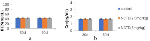
Figure 4. Effect of NCTD on kidney function of mice. (A, B) Even when the NCTD dose and application date were increased, the values of serum urea nitrogen and serum creatine did not change significantly.
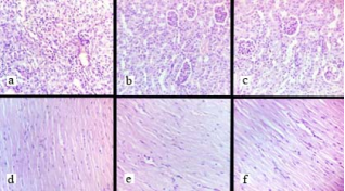
Figure 5. Effect of NCTD on kidney and myocardial tissue of mice. (H-E stain, 10×40) (a) Normal kidney tissue of mice injected with physiological saline on 60d. (b) Kidney tissue of mice injected with NCTD at 2.5mg/kg every day for 60d. (c) Kidney tissue of mice injected with NCTD at 5mg/kg every day for 60d. There was no significant difference in tubular epithelial cell degeneration and inflammatory cell infiltration compared to the control group injected with physiological saline after 60d injection of NCTD at 2.5mg/kg and 5mg/kg daily, and no bleeding glomeruli were found. (d) Normal myocardial tissue of mice injected with 60d physiological saline. (e) Myocardial tissue of mice injected with NCTD at 2.5mg/kg every day for 60d. (f) Myocardial tissue of mice injected with NCTD at 5mg/kg every day for 60d. After 60d injection of NCTD at 2.5mg/kg and 5mg/kg daily, the denatured myocardial cells and inflammatory cells were not significantly different from those injected with physiological saline.
3.3. Effect of NCTD on Hematopoietic Tissues of Mice
We performed peripheral blood examination and pathological biopsy of the spleen and bone marrow tissues to determine the effect of NCTD on the hematopoietic tissues of mice. The number of leucocytes and erythrocytes and the amount of hemoglobin were selected as the peripheral blood test index. When NCTD was individually injected at 2.5mg/kg and 5mg/kg per mouse daily for 30d or 60d, the changes in the peripheral blood according to the dose were observed. There were no significant differences in leuco- cytes, erythrocytes, and hemoglobin compared to the control (Figure 7-a, b). When NCTD was injected at a dose of 2.5mg/kg for 30d or 60d, the number of lymph nodes of spleen increased a bit, but When NCTD was injected at a dose of 5mg/kg for 30d or 60d, there were no significant differences in the number of them compared to the control (Figure 7-a, b, c). When NCTD is applied at a dose of 2.5mg/kg for 30d the rising tendency of the number of lymph node meant that this drug can give positive effect on immune system at a low concentration. During inoculation at a dose of 2.5mg/kg for 30d or 60d the volume of bone marrow was calculated in areal ratio and there was no difference in comparison while showing no appearance of adipose tissues in medulla cavity. (Figure 7-d, e, f)
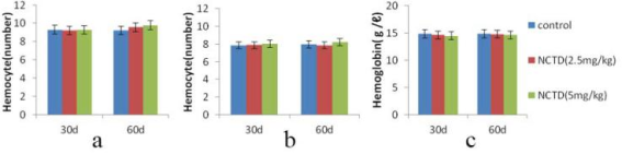
Figure 6. Effect of NCTD on peripheral blood test of mice. (a, b, c) Although the dose and application date of NCTD increases, there were no changes in the number of erythrocytes, leucocytes and hemoglobin.
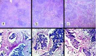
Figure 7. Effect of NCTD on spleen and marrow of mice. (H-E stain, spleen: 10×10, marrow: 10×40). (a) Normal spleen tissue of the mouse that was injected with physiological saline for 60d. (b) Spleen tissue of the mouse injected with NCTD at a dose of 2.5mg/kg daily for 60d. (c) Spleen tissue of the mouse injected with NCTD 5mg/kg daily for 60d. Injecting NCTD 2.5mg/kg daily for 60d, the number of lymph nodes increased a bit than the case of physiological saline and after injection of 5mg/kg, no big differences were found. (d) Normal marrow tissues injected with physiological saline for 60d. (e) Marrow tissues of the mouse injected with NCTD at a dose of 2.5mg/kg daily. (f) Marrow tissues of the mouse injected with NCTD 5mg/kg daily for 60d. The volume of marrow after injection of 2.5mg/kg, 5mg/kg daily for 60d shows no significant difference compared to the physiological saline.
4. Conclusion
NCTD exerts anticancer effects through mechanisms such as inhibition of cell proliferation, antiangiogenic effects, cell cycle blockade, and induction of cell apoptosis. Therefore, NCTD can be used for the treatment of primary liver cancer, esophageal cancer, breast cancer, gallbladder cancer, and leukemia .
In the treatment of liver cancer, high concentrations of NCTD (50-100 lm) are required for the treatment of apoptotic pathways in HCC cells, but the toxicity of NCTD is relatively high compared to normal hepatocytes. Also the effects of NCTD on the organs and tissues in vivo remain largely unknown.
We found that the acute toxicity of NCTD (LD50) was 20.75 mg/kg in the experiments. Also, we found that NCTD (2.5mg /kg or 5mg/kg per mouse for 5d) below a fourth concentration of LD50 had significant anti-angiogenesis effects in chamber- transplant models (Figure 1). On the basis of this experimental results we examined the effects of NCTD on the main organs and tissues of mice treated with 60 daily intraperitoneal injections.
Then injecting NCTD at a dose of 2.5 mg/kg for 30, 60d the value of ZTT did not increase compared to control group but at 5mg/kg there was significant increase in the value (Figure 2-C). Also, observing the morphological changes in the liver, after injecting NCTD at a dose of 5mg/kg for 30, 60d there was significant vacuolar degeneration in hepatocytes but at 2.5mg/kg for 30d with less degeneration. The inflammatory cells infiltrated into hepatic tissue as degenerated cells increased in number with more infiltration (Figure 3).
We found that NCTD had negative effect on liver as the dose and the application date increases but at the considerable dose (2.5 mg/kg) NCTD is harmless to the liver while having no negative effect on kidney and cardiac tissue. Also it is examined that it has positive influence on hematopoietic tissues after long period of application and especially it had tendency increasing the number of leucocytes. In another study, it was found that NCTD stimulated the cell cycle progression of granulocyte macrophage colony-forming cells (GM-CFC), DNA synthesis and the production of interleukin-1B, colony stimulating activity (CSA) and tumor necrosis factor (TNF)-α. .
Based on this study we suggest that unlike other chemical anti-cancer drugs, NCTD has no side effects on spleen and hematopoietic system and applying for a considerable period at a certain dose may not reduce the number of leucocyte but rather shows tendency of increasing which will have great influence on the onco-immune of the host. It is considered that NCTD is suitable for the application of the anti-cancer treatment and has available potential value to pre-surgical anti-cancer treatment.
Abbreviations

NCTD

Norcantharidin

MMP-9

Matrix Metallo-Proteinase-9

MRAP-2

Multidrug Resistance-Associated Protein 2

CTD

Cantharidin

NMR

NUCLEAR MAGNETIC RESONANCE

GM-CFC

Granulocyte Macrophage Colony-Forming Cells

CSA

Colony Stimulating Activity

TNF

Tumor Necrosis Factor

Author Contributions
Kwang-Il To: Project administration, Writing – review & editing
Dong-Min Han: Conceptualization, Formal analysis
Hong-Chol Ri: Conceptualization, Investigation, Data curation
Jong-Hyok Kim: Software, Formal analysis, Validation
Gyong-Jin Mun: Conceptualization, Formal analysis
Hui-Won Kim: Writing – original draft, Writing – review & editing, Investigation, Data curation.
Jun-hyok Ryang: Writing – original draft, Writing – review & editing, Investigation, Data curation
Chong-Hyok Han: Software, Formal analysis, Validation
Conflicts of Interest
The authors declare no conflicts of interest.
References
[1] A. Gritli-Linde, M. Bei, R. Maas, X. M. Zhang, A. Linde, and A. P. McMahon, Shh signaling within the dental epithelium is necessary for cell proliferation, growth and polarization, Development, 129(23), 5323–5337, 2002.
[2] Zhao, Jie; Guan, Xiao-Wen; Chen, Shi-Wu; Hui, Ling, Synthesis and biological evaluation of norcantharidin derivatives as protein phosphatase-1 inhibitors, Bioorganic and Medicinal Chemistry Letters, 25(2), 363-366, 2015.
[3] Sun, Hong; Li, Yifan; Li, Xinyu; Zhang, Yalin, The Inhibition of Serine/Threonine Protein Phosphatase Type 5 Mediates Cantharidin Toxicity to Control Periplaneta americana (L.), Insects, 11(10), 682-694, 2020.
[4] F. Cimmino, M. N. Scoppettuolo, M. Carotenuto et al., Norcantharidin impairs medulloblastoma growth by inhibition of Wnt/𝛽-catenin sign Journal of Neuro- Oncology, vol. 106, no. 1, pp. 59–70, 2012.
[5] Nohemí Percino-Daniel, David Buckley, Mario García-París, Pharmacological properties of blister beetles (Coleoptera: Meloidae) promoted their integration into the cultural heritage of native rural Spain as inferred by vernacular names diversity, traditions, and mitochondrial DNA, Journal of Ethnopharmacology, 147(3), 570-583, 2013.
[6] G. Turashvili, J. Bouchal, G. Burkadze, and Z. Kolar, Wnt signaling pathway in mammary gland development and carcinogenesis, Pathobiology, 73(5), 213–223, 2007.
[7] J. A. Sakoff, S. P. Ackland, M. L. Baldwin, M. A. Keane, and A. McCluskey, Anticancer activity and protein phosphatase 1 and 2A inhibition of a new generation of cantharidin analogues, Investigational New Drugs, 20(1), 1–11, 2002.
[8] Meng-Dan Xu, Lu Liu, Meng-Yao Wu, Min Jiang, Liu-Mei Shou et al., The combination of cantharidin and antiangiogenic therapeutics presents additive antitumor effects against pancreatic cancer, Oncogenesis, 7(94), 2018.
[9] K. A. Chuang, C. H. Lieu, W. J. Tsai et al., Evaluation of anti-Wnt/𝛽-catenin signaling agents by pGL4-TOP trans- fected stable cells with a luciferase reporter system, Brazilian Journal of Medical and Biological Research, 43(10), 931–941, 2010.
[10] L. Yang, G. Xie, Q. Fan, and J. Xie, Activation of the hedgehog signaling pathway in human cancer and the clinical implications, Oncogene, 29(4), 469–481, 2010.
[11] M. Guan, Q. L. Zhu, Y. Liu et al., Uptake and transport of a novel anticancer drug-delivery system: lactosyl norcantharidin -associated N-trimethyl chitosan nanoparticles across intestinal Caco-2 cell monolayers, International Journal of Nanomedicine, 7, 1921–1930, 2012.
[12] Junran Xie, Yaping Zhang, Xuming Hu, Ran Lv, Dongju Xiao, Li Jiang and Qi Bao, Norcantharidin inhibits Wnt signal pathway via promoter demethylation of WIF-1 in human non-small cell lung cancer, Medical Oncology, 32(145), 1-7, 2015.
[13] P.-Y. Yang, M.-F. Chen, Y.-H. Kao, D.-N. Hu, F.-R. Chang, and Y.-C. Wu, Norcantharidin induces apoptosis of breast cancer cells: involvement of activities of mitogen activated protein kinases and signal transducers and activators of transcription, Toxicology in Vitro, 25(3), 699–707, 2011.
[14] R. L. Carpenter and H. W. Lo, Hedgehog pathway and GLI1 isoforms in human cancer, Discovery Medicine, 13(69), 105–113, 2012.
[15] S.-H. Kok, S.-J. Cheng, C.-Y. Hong et al., Norcantharidin induced apoptosis in oral cancer cells is associated with an increase of proapoptotic to anti-apoptotic protein ratio, Cancer Letters, 217(1), 43–52, 2005.
[16] S. V. Ambudkar, C. Kimchi-Sarfaty, Z. E. Sauna, and M. M. Gottesman, P-glycoprotein: from genomics to mechanism, Oncogene, 22(47), 7468–7485, 2003.
[17] Chu-Liang Lin, Chien-Min Chen, Chia-Liang Lin et al., Norcantharidin induces mitochondrial-dependent apoptosis through Mcl-1 inhibition in human prostate cancer cell, Biochimica et Biophysica Acta (BBA) - Molecular Cell Research, 10(1864), 1867-1876, 2017.
[18] Y.-C. Chen, S.-C. Chang, M.-H. Wu et al., “Norcantharidin reduced cyclins and cytokines production in human peripheral blood mononuclear cells,” Life Sciences, vol. 84, no. 7-8, pp. 218–226, 2009.
[19] Chen-Hsi Hsieh, K. S. Clifford Chao, Hui-Fen Liao and Yu-Jen Chen, Norcantharidin, Derivative of Cantharidin, for Cancer Stem Cells, Evidence-Based Complementary and Alternative Medicine, 838651, 1-11, 2013.
[20] Fanqin Zhang, Xiaodong Chen, Chuanqi Qiao, et al, Exploring the Anti-Colorectal Cancer Mechanism of Norcantharidin Through TRAF5/NF-κB Pathway Regulation and Folate-Targeted Liposomal Delivery, International Journal of Molecular Sciences, 26, 1450, 2025.
[21] Zeping Han, Baoxia Li, Juanjuan Wang, Xiangqiang Zhang, “Norcantharidin Inhibits SK-N-SH Neuroblastoma Cell Growth by Induction of Autophagy and Apoptosis”, Technology in Cancer Research & Treatment, 1(16), 2016.
[22] Y.-J. Chen, W.-M. Chang, Y.-W. Liu et al., A small-molecule metastasis inhibitor, norcantharidin, downregulates matrix metalloproteinase-9 expression by inhibiting Sp1 transcriptional activity in colorectal cancer cells, Chemico-Biological Interactions, 181(3), 440–446, 2009.
[23] Y.-J. Chen, Y.-M. Tsai, C.-D. Kuo, K.-L. Ku, H.-S. Shie, and H.-F. Liao, Norcantharidin is a small-molecule synthetic com- pound with anti-angiogenesis effect, Life Sciences, 85(17), 642–651, 2009.
[24] Y.-N. Chen, C.-C. Cheng, J.-C. Chen, W. Tsauer, and S.-L. Hsu, Norcantharidin-induced apoptosis is via the extracellular signal-regulated kinase and c-Jun-NH2-terminal kinase signal- ing pathways in human hepatoma HepG2 cells, British Journal of Pharmacology, 140(3), 461–470, 2003.
[25] Y. Y. Bei, X. Y. Chen, Y. Liu et al., Novel norcantharidin-loaded liver targeting chitosan nanoparticles to enhance intestinal absorption, International Journal of Nanomedicine, 7, 1819–1827, 2012.
[26] Y.-Z. Fan, J.-Y. Fu, Z.-M. Zhao, and C.-Q. Chen, Effect of norcantharidin on proliferation and invasion of human gallbladder carcinoma GBC-SD cells, World Journal of Gastroenterology, 11(16), 2431-2437, 2005.
[27] Jiwei Guo, Yan Wu, Jing Du, Lijuan Yang et al., Deregulation of UBE2C-mediated autophagy repression aggravates NSCLC progression, Oncogenesis, 7(49), 2018.
[28] Y.-Z. Fan, Z.-M. Zhao, J.-Y. Fu, C.-Q. Chen, and W. Sun, Norcantharidin inhibits growth of human gallbladder carcinoma xenografted tumors in nude mice by inducing apoptosis and blocking the cell cycle in vivo, Hepatobiliary and Pancreatic Diseases International, 9(4), 414–422, 2010.
Cite This Article
  • APA Style

    To, K., Han, D., Ri, H., Kim, J., Mun, G., et al. (2025). Effects of Norcantharidin on the Main Organs and Tissues of Mice. Advances, 6(4), 123-130. https://doi.org/10.11648/j.advances.20250604.13

    Copy | Download

    ACS Style

    To, K.; Han, D.; Ri, H.; Kim, J.; Mun, G., et al. Effects of Norcantharidin on the Main Organs and Tissues of Mice. Advances. 2025, 6(4), 123-130. doi: 10.11648/j.advances.20250604.13

    Copy | Download

    AMA Style

    To K, Han D, Ri H, Kim J, Mun G, et al. Effects of Norcantharidin on the Main Organs and Tissues of Mice. Advances. 2025;6(4):123-130. doi: 10.11648/j.advances.20250604.13

    Copy | Download

  • @article{10.11648/j.advances.20250604.13,
      author = {Kwang-Il To and Dong-Min Han and Hong-Chol Ri and Jong-Hyok Kim and Gyong-Jin Mun and Hui-Won Kim and Jun-hyok Ryang and Chong-Hyok Han},
      title = {Effects of Norcantharidin on the Main Organs and Tissues of Mice},
      journal = {Advances},
      volume = {6},
      number = {4},
      pages = {123-130},
      doi = {10.11648/j.advances.20250604.13},
      url = {https://doi.org/10.11648/j.advances.20250604.13},
      eprint = {https://article.sciencepublishinggroup.com/pdf/10.11648.j.advances.20250604.13},
      abstract = {Norcantharidin (NCTD), a demethylated derivative of cantharidin, reportedly exhibits various biological anticancer activities including apoptosis, inhibition of cell proliferation, cell-cycle blockage, induction of cell apoptosis, and anti-angiogenesis. NCTD is currently used as an anticancer drug for hepatoma, breast cancer and colorectal adenocarcinoma. However, the effects of NCTD on the organs and tissues in vivo remain largely unknown. Here, we pathologically examined the effects of NCTD on the organs and tissues including liver, heart, kidney, spleen and medulla in mice. After mice were treated with 60 daily intraperitoneal injections of NCTD (2.5 mg/kg or 5 mg/kg), toxicity on the heart and kidneys were not discovered but on the liver was a significant increase according to the injection dose and the date. The number of leucocytes had a tendency to rise in the peripheral blood test and there weren’t any pathological changes in the spleen and medulla. NCTD has no side effects on spleen and hematopoietic system and applying for a considerable period at a certain dose may not reduce the number of leucocyte but rather shows tendency of increasing which will have great influence on the onco-immune of the host. It is concluded that NCTD is suitable for the application of the anti-cancer treatment with no myelo-suppression.},
     year = {2025}
    }
    

    Copy | Download

  • TY  - JOUR
    T1  - Effects of Norcantharidin on the Main Organs and Tissues of Mice
    AU  - Kwang-Il To
    AU  - Dong-Min Han
    AU  - Hong-Chol Ri
    AU  - Jong-Hyok Kim
    AU  - Gyong-Jin Mun
    AU  - Hui-Won Kim
    AU  - Jun-hyok Ryang
    AU  - Chong-Hyok Han
    Y1  - 2025/12/24
    PY  - 2025
    N1  - https://doi.org/10.11648/j.advances.20250604.13
    DO  - 10.11648/j.advances.20250604.13
    T2  - Advances
    JF  - Advances
    JO  - Advances
    SP  - 123
    EP  - 130
    PB  - Science Publishing Group
    SN  - 2994-7200
    UR  - https://doi.org/10.11648/j.advances.20250604.13
    AB  - Norcantharidin (NCTD), a demethylated derivative of cantharidin, reportedly exhibits various biological anticancer activities including apoptosis, inhibition of cell proliferation, cell-cycle blockage, induction of cell apoptosis, and anti-angiogenesis. NCTD is currently used as an anticancer drug for hepatoma, breast cancer and colorectal adenocarcinoma. However, the effects of NCTD on the organs and tissues in vivo remain largely unknown. Here, we pathologically examined the effects of NCTD on the organs and tissues including liver, heart, kidney, spleen and medulla in mice. After mice were treated with 60 daily intraperitoneal injections of NCTD (2.5 mg/kg or 5 mg/kg), toxicity on the heart and kidneys were not discovered but on the liver was a significant increase according to the injection dose and the date. The number of leucocytes had a tendency to rise in the peripheral blood test and there weren’t any pathological changes in the spleen and medulla. NCTD has no side effects on spleen and hematopoietic system and applying for a considerable period at a certain dose may not reduce the number of leucocyte but rather shows tendency of increasing which will have great influence on the onco-immune of the host. It is concluded that NCTD is suitable for the application of the anti-cancer treatment with no myelo-suppression.
    VL  - 6
    IS  - 4
    ER  - 

    Copy | Download

Author Information
  • Institute of Chemistry and Biology, University of Sciences, Pyongyang, DPR Korea

  • Institute of Chemistry and Biology, University of Sciences, Pyongyang, DPR Korea

  • Department of Pathology, Pyongyang University of Medical Sciences, Pyongyang, DPR Korea

  • Institute of Chemistry and Biology, University of Sciences, Pyongyang, DPR Korea

  • Institute of Chemistry and Biology, University of Sciences, Pyongyang, DPR Korea

  • Institute of Microbiology, State Academy of Science, Pyongyang, DPR Korea

  • Institute of Microbiology, State Academy of Science, Pyongyang, DPR Korea

  • Clinical Faculty No 3., Pyongsong Medical College, Pyongsong, DPR Korea